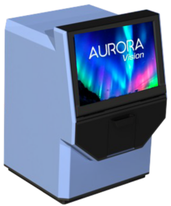
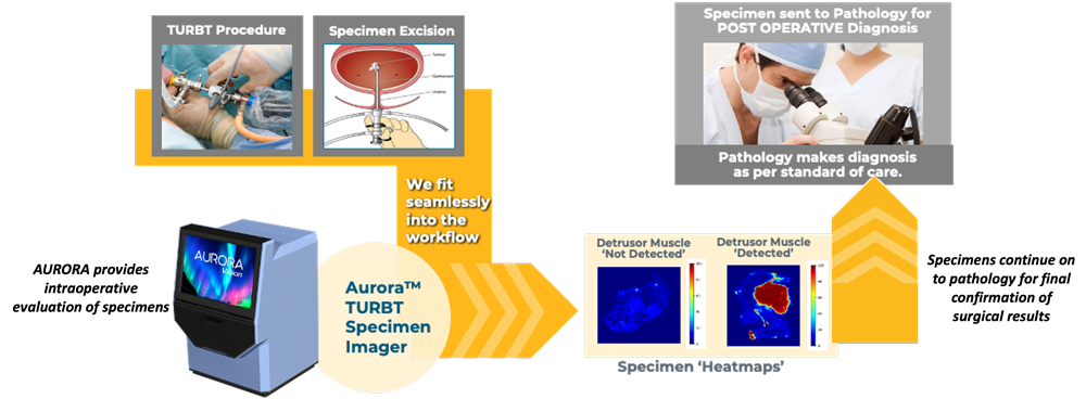
The AURORA™ TURBT Specimen Imager based on CytoVeris’ Multi-Spectral Autofluorescence imaging technology, is designed for normal tissue discrimination and is powered by our tissue classification AI-based algorithmAURORAVision
AURORA, a bench top unit is designed for small specimen analysis: 10mm dia, and as such is suitable for biopsy specimen analysis. We have launched Aurora targeted for use in non-muscle invasive (NMI) bladder cancer and the analysis of specimens in transurethral resection of bladder tumor (TURBT) procedures.
Real-time imaging during surgery could improve the odds of preserving function and reducing recurrence.
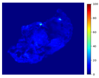
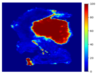
Specimen ‘Heatmaps’
Detrusor Muscle ‘Not Detected’
Detrusor Muscle ‘Not Detected’
Detrusor Muscle ‘Detected’
Detrusor Muscle ‘Detected’
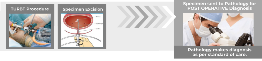
In TURBT surgeries, to allow for the correct staging of the cancer, it is essential that the surgeon is able to resect tissue to the outer bladder ‘detrusor’ muscle wall.
The AURORAVision outputs above indicate the application of the device ‘trained’ to detect DM in specimens. The output can be displayed as a heatmap or binary output
Thecurrent standard of care in TURBT
AURORAfits seamlessly into the TURBT surgical workflow