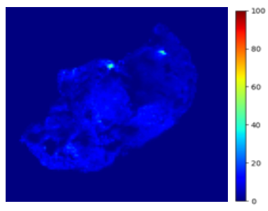
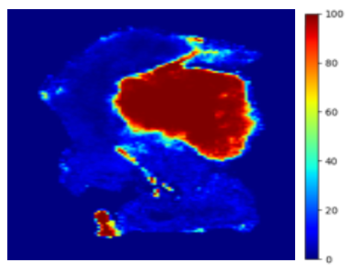
Detection of Intrinsic Tissue Biomolecular Identifiers using Aurora Visions
The Aurora Tissue Imaging system is a benchtop device into which excised specimens are placed and imaged. The system has an intuitive AI produced output that displays the presence/absence of target or cancerous tissue to help guide the surgeon’s decision-making. Aurora does not disrupt the established workflow or standard of care or significantly extend OR time.
The Aurora Tissue Imaging System fits seamlessly into the TURBT clinical workflow
TURBT
Procedure
>
Specimen Excision
>
No need to send the sample out of the OR to Pathology for POST OPERATIVE Diagnosis where the Pathologist makes diagnosis as per standard of care.
>
Real-time Imaging with no disruption
Detection of using Autofluorescence
STEP 1
Multi-Spectral Illumination
STEP 2
Multi-Spectral Detection
STEP 3
Train AI/ML Algorithms
Specimen ‘Heatmaps’
Real-time imaging during surgery could improve the odds of preserving function and reducing recurrence.
Detrusor Muscle ‘Not Detected’
Detrusor Muscle ‘Detected’